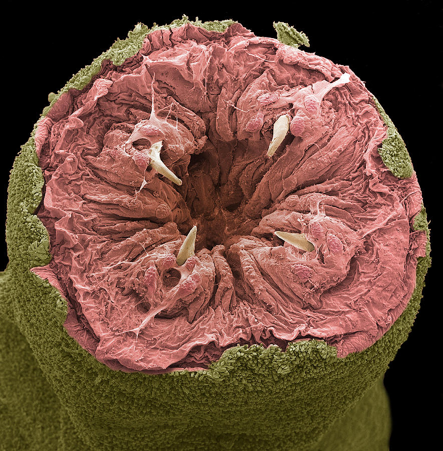
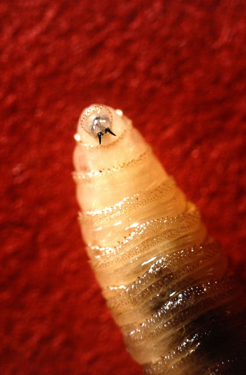
"Worms in mouth pictures" refers to a medical condition known as myiasis, where fly larvae infest and feed on the tissues of a living organism. These larvae can infest the mouth, causing significant damage and discomfort to the affected individual. Myiasis is a serious condition and requires prompt medical attention to prevent further complications.
Myiasis can occur when fly eggs are deposited in open wounds, skin folds, or body orifices. The larvae hatch and begin to feed on the surrounding tissue, causing pain, swelling, and discharge. In the case of oral myiasis, the larvae can infest the gums, tongue, cheeks, or palate, leading to extensive tissue damage and potential tooth loss.
Treatment for oral myiasis typically involves removing the larvae and administering antibiotics to prevent infection. In severe cases, surgery may be necessary to remove damaged tissue and restore oral function. Prevention of myiasis involves avoiding contact with flies, keeping wounds clean and covered, and promptly addressing any skin or oral infections.
Worms in Mouth Pictures
Worms in mouth pictures, also known as oral myiasis, is a serious medical condition caused by fly larvae infesting the mouth. Understanding the various aspects of this condition is crucial for effective diagnosis, treatment, and prevention.
- Definition: Infestation of mouth tissues by fly larvae.
- Causes: Deposition of fly eggs in open wounds or body orifices.
- Symptoms: Pain, swelling, discharge, and tissue damage.
- Treatment: Removal of larvae and antibiotics.
- Prevention: Avoiding contact with flies and keeping wounds clean.
- Complications: Extensive tissue damage and tooth loss.
- Diagnosis: Visual examination and identification of larvae.
- Epidemiology: More common in tropical and subtropical regions.
- Public Health: Can be associated with poor sanitation and hygiene.
- Research: Ongoing studies on larval biology and treatment options.
In conclusion, worms in mouth pictures, or oral myiasis, is a significant medical condition that requires prompt attention. Understanding the key aspects discussed above, including causes, symptoms, treatment, and prevention, is essential for effective management of this condition. Further research and public health initiatives are needed to address the underlying factors contributing to oral myiasis and improve outcomes for affected individuals.
Definition
This definition captures the essence of "worms in mouth pictures," highlighting the presence of fly larvae within the mouth's soft tissues. Understanding this definition is crucial for comprehending the condition's nature and implications.
- Pathophysiology: Fly larvae require a moist environment to survive and feed, making the mouth an ideal location for infestation. They can penetrate the oral mucosa, causing significant inflammation and tissue damage.
- Clinical Manifestations: Patients with oral myiasis may experience severe pain, swelling, and discharge from the mouth. The larvae can invade the gums, tongue, cheeks, and palate, leading to extensive tissue destruction and potential tooth loss.
- Diagnosis: Visual examination by a healthcare professional is essential for diagnosing oral myiasis. The presence of white or cream-colored larvae within the mouth confirms the diagnosis.
- Treatment: Prompt removal of the larvae is crucial to prevent further tissue damage and infection. This can be achieved through mechanical extraction or irrigation with antiseptic solutions. Antibiotics are often prescribed to combat any secondary bacterial infections.
In summary, understanding the definition of "infestation of mouth tissues by fly larvae" is fundamental to recognizing and managing "worms in mouth pictures." This definition underscores the presence of fly larvae within the oral cavity, leading to a cascade of pathological, clinical, diagnostic, and therapeutic considerations.
Causes
The deposition of fly eggs in open wounds or body orifices is directly linked to the development of "worms in mouth pictures," also known as oral myiasis. Understanding this connection is vital for effective prevention and management of this condition.
When flies lay their eggs in open wounds or body orifices, the larvae that hatch from these eggs can invade and infest the surrounding tissues. In the case of oral myiasis, the larvae can enter the mouth through open wounds, such as surgical incisions or dental extractions, or through natural orifices like the nose or eyes. Once inside the mouth, the larvae feed on the soft tissues, causing significant damage and discomfort.
Preventing the deposition of fly eggs in open wounds or body orifices is crucial to reducing the risk of oral myiasis. This involves maintaining good hygiene, promptly addressing any wounds or injuries, and using insect repellent when necessary. Additionally, proper waste management and sanitation practices can help reduce fly populations and further minimize the risk of infestation.
In conclusion, understanding the connection between the deposition of fly eggs in open wounds or body orifices and "worms in mouth pictures" is essential for developing effective preventive measures and ensuring timely treatment. By implementing appropriate hygiene and sanitation practices, individuals can significantly reduce their risk of developing this potentially serious condition.
Symptoms
The symptoms of "worms in mouth pictures," or oral myiasis, are directly linked to the presence and activity of fly larvae within the oral cavity. Understanding this connection is crucial for accurate diagnosis and effective management of this condition.
The feeding activity of fly larvae causes significant damage to the soft tissues of the mouth, leading to a range of symptoms. Pain, swelling, and discharge are common manifestations of oral myiasis. The pain can be severe and persistent, interfering with daily activities such as eating and speaking. Swelling can cause difficulty opening the mouth and may extend to the face and neck. Discharge from the mouth may be purulent or blood-tinged, indicating the presence of infection.
In severe cases, the larvae can cause extensive tissue damage, leading to the destruction of oral structures. This can result in tooth loss, damage to the palate or tongue, and even bone erosion. The presence of larvae and the associated tissue damage can also create a favorable environment for bacterial growth, increasing the risk of secondary infections.
Recognizing the symptoms of oral myiasis and seeking prompt medical attention is essential to prevent complications and ensure effective treatment. Accurate diagnosis and timely intervention can help minimize tissue damage, reduce pain and discomfort, and prevent the spread of infection.
Treatment
In the context of "worms in mouth pictures," or oral myiasis, the treatment involves a two-pronged approach centered around the removal of fly larvae and the administration of antibiotics. Understanding this connection is pivotal for effective management and successful resolution of this condition.
- Removal of Larvae:
The primary goal of treatment is the meticulous removal of fly larvae from the oral cavity. This can be achieved through various techniques, including manual extraction using forceps or irrigation with antiseptic solutions. Thorough removal of all larvae is crucial to prevent further tissue damage and eliminate the source of infection.
- Antibiotics:
Antibiotics play a crucial role in combating secondary bacterial infections that often accompany oral myiasis. The presence of larvae creates an environment conducive to bacterial growth, increasing the risk of complications. Antibiotics are prescribed to target and eliminate these bacterial infections, preventing their spread and further damage to the oral tissues.
The combination of larval removal and antibiotic therapy is essential for effective treatment of oral myiasis. Prompt intervention and proper execution of these measures can significantly improve outcomes, reduce the severity of symptoms, and prevent long-term complications.
Prevention
In the context of "worms in mouth pictures," also known as oral myiasis, prevention is of paramount importance to mitigate the risk of infestation and its associated complications. Understanding the connection between "Prevention: Avoiding contact with flies and keeping wounds clean" and oral myiasis is crucial for effective self-care and public health measures.
- Avoiding Contact with Flies:
Flies are the primary vectors responsible for transmitting oral myiasis. Limiting contact with flies reduces the likelihood of egg deposition in open wounds or body orifices, which is the initial step in the development of oral myiasis. Simple measures such as using insect repellent, wearing protective clothing, and maintaining a clean environment can significantly reduce fly exposure and the risk of infestation.
- Keeping Wounds Clean:
Open wounds provide an ideal environment for fly egg deposition and subsequent larval infestation. Proper wound care, including thorough cleaning, regular dressing changes, and avoiding exposure to unsanitary conditions, is essential in preventing oral myiasis. Ensuring prompt medical attention for any wounds, especially those involving the mouth, is crucial to minimize the risk of infection and potential infestation.
By implementing these preventive measures, individuals can significantly reduce their chances of developing oral myiasis. Public health initiatives aimed at improving sanitation, waste management, and fly control can further contribute to reducing the prevalence of this condition, especially in vulnerable populations and resource-limited settings.
Complications
Within the context of "worms in mouth pictures," or oral myiasis, complications arise from the destructive nature of the fly larvae infesting the oral cavity. Understanding the connection between "Complications: Extensive tissue damage and tooth loss" and oral myiasis is crucial for recognizing the potential severity and impact of this condition.
The feeding activity of fly larvae within the mouth causes significant tissue damage, leading to a range of complications. The larvae can penetrate and erode the soft tissues of the mouth, including the gums, tongue, cheeks, and palate. This damage can result in pain, swelling, and difficulty eating and speaking. In severe cases, the larvae can cause extensive tissue destruction, leading to tooth loss and even bone erosion.
Beyond the immediate damage caused by the larvae, oral myiasis can also lead to secondary infections due to the presence of bacteria and the breakdown of tissues. These infections can further contribute to tissue damage and tooth loss. Additionally, the presence of larvae and the associated inflammation can interfere with the normal healing process, making it more challenging for the mouth to repair itself.
Recognizing the potential complications of oral myiasis is essential for prompt diagnosis and appropriate treatment. Early intervention can help minimize tissue damage, reduce the risk of tooth loss, and prevent the development of secondary infections. Proper wound care and antibiotic therapy are crucial in managing oral myiasis and preventing complications.
Diagnosis
In the context of "worms in mouth pictures," or oral myiasis, accurate diagnosis is crucial for effective treatment and prevention of complications. Visual examination and identification of larvae play a central role in the diagnostic process, providing direct evidence of the infestation and guiding appropriate management.
- Visual Examination:
A thorough visual examination of the oral cavity is essential for diagnosing oral myiasis. Healthcare professionals use specialized instruments to examine the mouth, paying close attention to areas where larvae are commonly found, such as the gums, tongue, and palate. The presence of white or cream-colored larvae, along with signs of inflammation and tissue damage, strongly suggests oral myiasis.
- Identification of Larvae:
Once larvae are visualized, their identification is crucial for confirming the diagnosis and determining the appropriate treatment. Healthcare professionals may collect samples of the larvae for microscopic examination or consult with entomologists for expert identification. Accurate identification of the larvae helps guide decisions regarding specific medications and management strategies.
By combining visual examination with careful identification of larvae, healthcare professionals can accurately diagnose oral myiasis, enabling prompt intervention and appropriate treatment to minimize tissue damage and prevent complications. This underscores the critical role of visual examination and identification of larvae in managing "worms in mouth pictures" and safeguarding oral health.
Epidemiology
Understanding the connection between the epidemiology of "worms in mouth pictures," also known as oral myiasis, and its prevalence in tropical and subtropical regions is crucial for effective prevention and control strategies. The geographic distribution of this condition is influenced by a complex interplay of environmental, socioeconomic, and behavioral factors.
In tropical and subtropical regions, the warm and humid climate provides an ideal environment for the flies that transmit oral myiasis to thrive and reproduce. These regions often have high populations of livestock, which attract flies and increase the risk of human exposure to fly larvae. Additionally, poor sanitation and hygiene practices in some tropical and subtropical areas contribute to the proliferation of flies and the spread of oral myiasis.
Understanding the epidemiology of oral myiasis is essential for public health interventions aimed at reducing its incidence and impact. Targeted control measures, such as vector control, improved sanitation, and health education, can be implemented to mitigate the risk of oral myiasis in vulnerable populations.
Recognizing the geographic distribution and epidemiological factors associated with oral myiasis is crucial for healthcare professionals, policymakers, and individuals living in affected areas. This knowledge empowers them to take appropriate preventive measures, seek timely diagnosis and treatment, and contribute to reducing the burden of this condition.
Public Health
The connection between public health and "worms in mouth pictures," or oral myiasis, highlights the significant impact of sanitation and hygiene practices on the prevalence and prevention of this condition. Poor sanitation and hygiene create favorable conditions for the transmission and development of oral myiasis, posing a public health challenge in many regions.
- Environmental Factors:
Lack of access to clean water, inadequate waste management, and poor sanitation practices contribute to the proliferation of flies, the vectors of oral myiasis. These unsanitary conditions provide breeding grounds for flies, increasing the risk of human exposure to their larvae.
- Personal Hygiene:
Poor oral hygiene, including infrequent brushing and flossing, can create an environment conducive to the development of oral myiasis. Food debris and bacteria in the mouth attract flies and increase the likelihood of egg deposition in open wounds or lesions.
- Healthcare Access:
Limited access to healthcare services, particularly in underserved communities, can delay diagnosis and treatment of oral myiasis. This delay can lead to severe complications, including extensive tissue damage and tooth loss.
- Education and Awareness:
Lack of education and awareness about oral myiasis and its prevention can contribute to its persistence. Public health campaigns and educational initiatives are crucial for informing communities about the importance of proper sanitation, hygiene, and prompt medical attention.
Addressing the public health aspects of oral myiasis requires a multi-faceted approach involving improvements in sanitation, hygiene practices, healthcare accessibility, and education. By promoting good public health practices, we can significantly reduce the incidence of oral myiasis and protect the health of vulnerable populations.
Research
Ongoing research plays a vital role in advancing our understanding and management of "worms in mouth pictures," also known as oral myiasis. Scientists are actively engaged in studying the biology of fly larvae and exploring novel treatment approaches to improve outcomes for affected individuals.
- Larval Biology and Identification:
Researchers are investigating the life cycle, behavior, and genetic diversity of fly larvae that cause oral myiasis. This knowledge is crucial for developing targeted interventions and improving diagnostic techniques to identify the specific species involved.
- Larval Pathogenicity and Host Response:
Studies focus on understanding the mechanisms by which fly larvae cause tissue damage and elicit host immune responses. This research aims to identify potential therapeutic targets and develop strategies to mitigate the adverse effects of larval infestation.
- Novel Treatment Approaches:
Researchers are exploring alternative treatment modalities, such as laser therapy and photodynamic therapy, to eliminate fly larvae and minimize tissue damage. These approaches aim to reduce the reliance on traditional surgical removal and antibiotics, potentially improving patient outcomes.
- Drug Development and Resistance Monitoring:
Ongoing research involves evaluating the efficacy and safety of new drugs and antibiotics against fly larvae. Additionally, studies monitor the development of resistance to existing treatments, ensuring the continued effectiveness of therapeutic interventions.
These research endeavors contribute significantly to the advancement of oral myiasis management. By gaining a deeper understanding of larval biology and treatment options, healthcare professionals can provide more effective and personalized care for patients affected by "worms in mouth pictures."
Frequently Asked Questions about "Worms in Mouth Pictures"
This section addresses common concerns and misconceptions surrounding oral myiasis, also known as "worms in mouth pictures," providing concise and evidence-based answers.
Question 1: How do fly larvae get into the mouth?
Fly larvae can enter the mouth through open wounds, such as surgical incisions or dental extractions, or through natural orifices like the nose or eyes.
Question 2: What are the symptoms of oral myiasis?
Symptoms include pain, swelling, discharge, and tissue damage in the mouth. The presence of white or cream-colored larvae is a telltale sign of infestation.
Question 3: How is oral myiasis treated?
Treatment involves removing the larvae and administering antibiotics to prevent or treat secondary infections.
Question 4: What are the potential complications of oral myiasis?
Complications include extensive tissue damage, tooth loss, and secondary infections, which can lead to severe health consequences if left untreated.
Question 5: How can oral myiasis be prevented?
Preventive measures include avoiding contact with flies, keeping wounds clean, and practicing good oral hygiene.
Question 6: Is oral myiasis common?
Oral myiasis is more common in tropical and subtropical regions where flies are prevalent. However, it can occur anywhere if properare not followed.
Summary: Oral myiasis is a serious condition that requires prompt medical attention. Understanding its causes, symptoms, treatment, and preventive measures is crucial for maintaining good oral health and preventing complications.
Transition to the next article section: For more information on oral myiasis, including research advancements and public health implications, please refer to the following sections.
Tips Regarding "Worms in Mouth Pictures"
To effectively manage and prevent oral myiasis, commonly known as "worms in mouth pictures," consider implementing the following evidence-based tips:
Tip 1: Maintain Good Oral Hygiene: Regularly brush and floss your teeth to remove food debris and bacteria that attract flies. This practice helps create an environment less conducive to larval development.
Tip 2: Protect Open Wounds: Keep wounds clean and covered to prevent fly exposure and egg deposition. If a wound becomes infected, seek medical attention promptly.
Tip 3: Avoid Contact with Flies: Use insect repellent, wear protective clothing, and keep your environment clean to minimize fly exposure and reduce the risk of infestation.
Tip 4: Practice Proper Sanitation: Ensure proper waste disposal and maintain a clean living environment to reduce fly populations and the likelihood of oral myiasis transmission.
Tip 5: Seek Medical Attention: If you suspect oral myiasis, seek medical attention immediately. Early diagnosis and treatment can minimize tissue damage and prevent complications.
Summary: By following these tips, you can significantly reduce your risk of developing oral myiasis and maintain good oral health. Remember, prevention is key, and prompt medical attention is crucial if an infestation occurs.
Transition to the article's conclusion: Understanding the importance of these tips empowers you to take proactive measures against oral myiasis, safeguarding your oral health and overall well-being.
Conclusion
Oral myiasis, commonly referred to as "worms in mouth pictures," is a serious medical condition caused by fly larvae infesting the mouth. Understanding its causes, symptoms, treatment, and preventive measures is crucial for maintaining good oral health and preventing complications. While oral myiasis is more prevalent in tropical and subtropical regions, it can occur anywhere if proper hygiene practices are not followed.
This article has explored the various aspects of oral myiasis, emphasizing the importance of early diagnosis, prompt treatment, and effective prevention. By implementing the tips outlined in the previous section, individuals can significantly reduce their risk of developing this condition and protect their oral health. Additionally, ongoing research and public health initiatives are essential for advancing our understanding and management of oral myiasis, ultimately improving outcomes for affected individuals.
Unraveling The Secrets Of "That's The Point With Brandon"
Unveiling The Profound Power Of Dancing Without Leaving Room For Jesus
Unveiling The Profound Meaning Behind "Tatuaje Rosario En La Mano"

Ragworm Mouth, Sem Photograph by Steve Gschmeissner

Worm Free Stock Photo Tusklike mandibles protruding from the
ncG1vNJzZmigla3Bs62NmqSsa16Ztqi105qjqJuVlru0vMCcnKxmk6S6cMPOq6SsZZmjeq671K2fZqiZmMG2vsSsZaGsnaE%3D